Featured Inventory
Your Source for the Microscopy Equipment and Electron Microscopes You Need
At J. Kraft Microscopy, we are focused on providing our customers with the best new and reconditioned equipment available. Thanks to our extensive network of industry partners and vast inventory across a variety of brands, we can best match the right products to your application.
To Meet our customers unique needs, our product offerings go beyond our standard inventory. We will work with you to configure custom systems, instrumentation, and parts to give you exactly what you need.
If you have question or are uncertain about what product will meet you needs best, you can request more information through our microscopy products information form. We are here to help provide you with the information you need, to make the best purchasing decision.
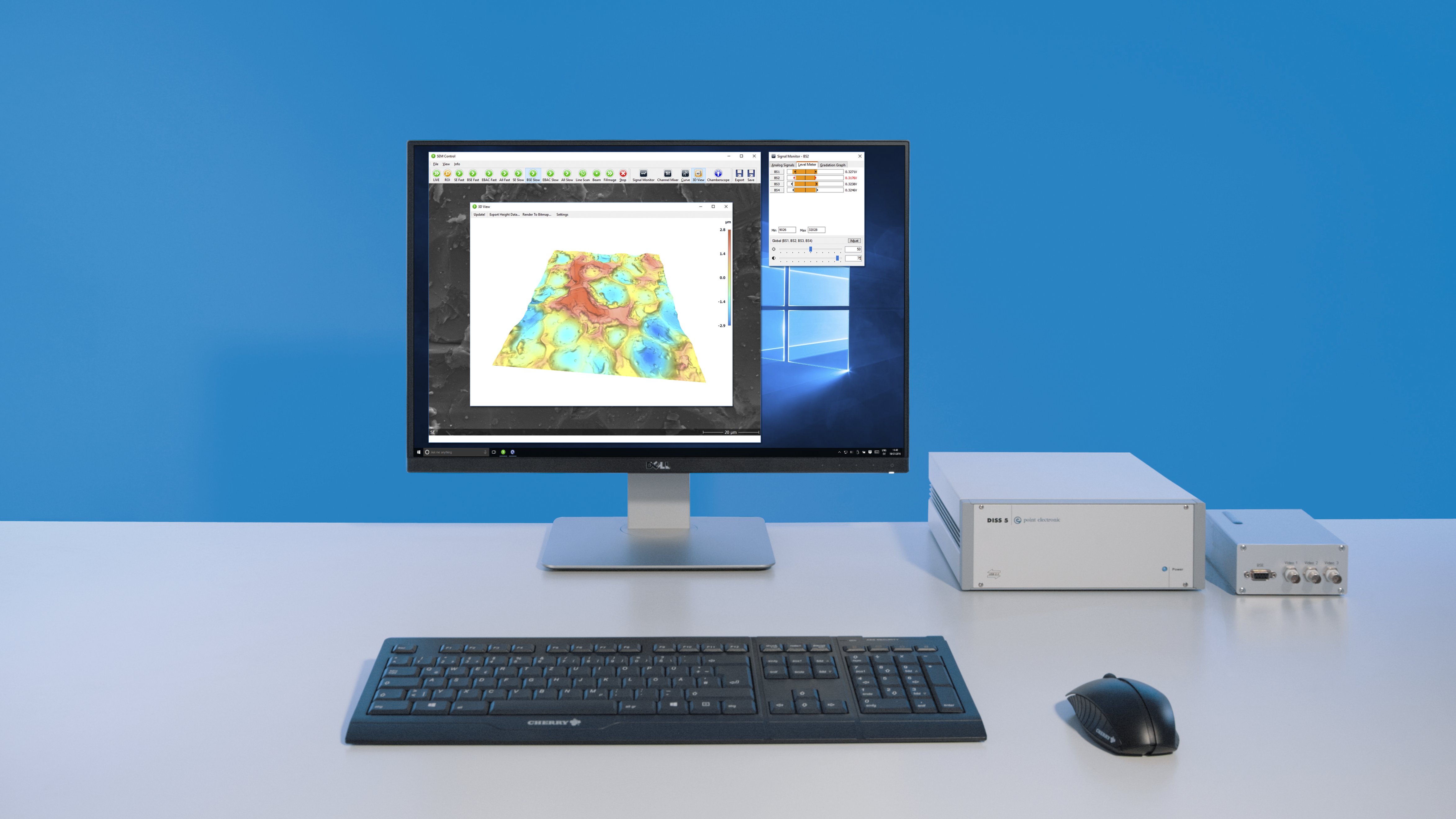
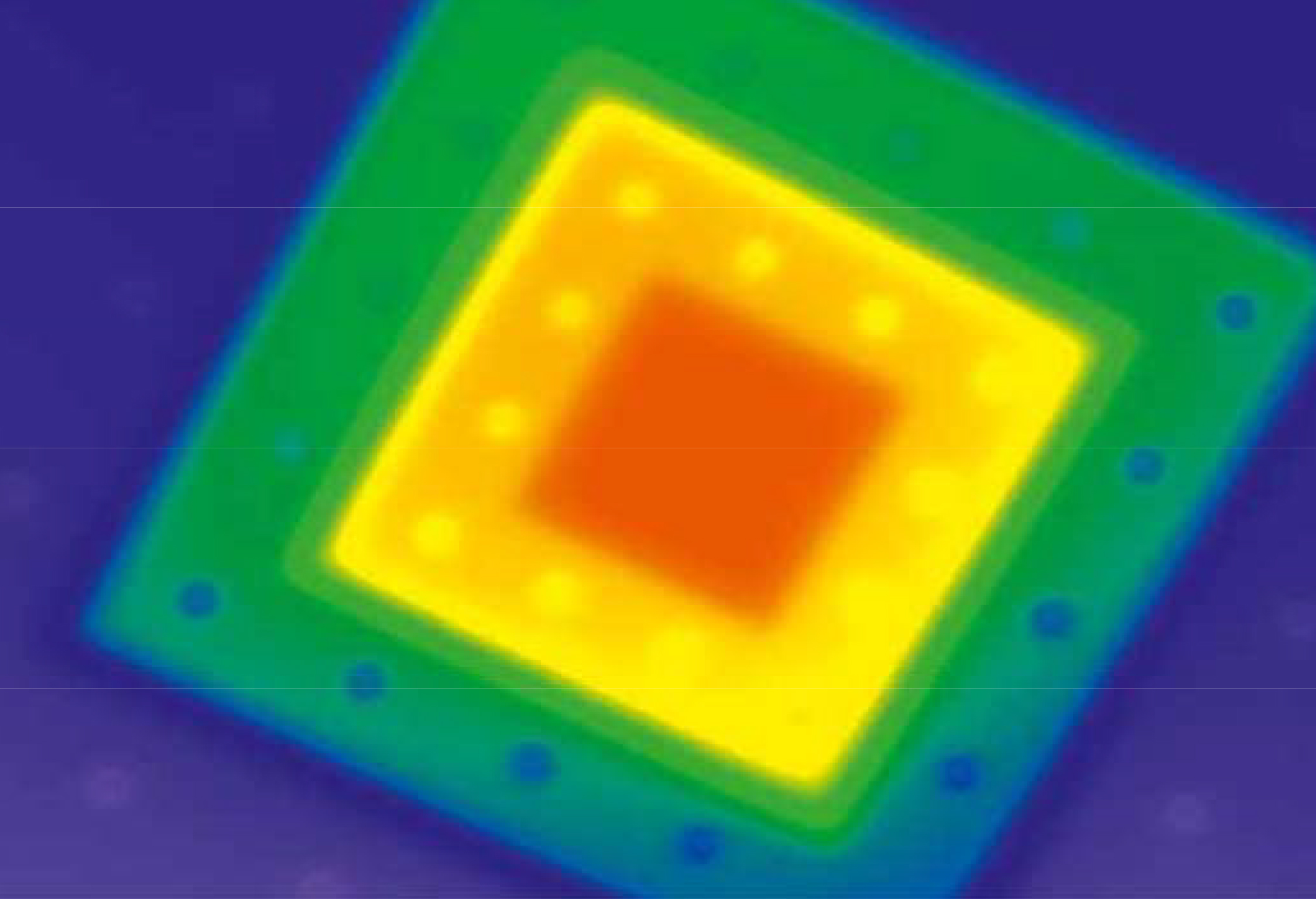
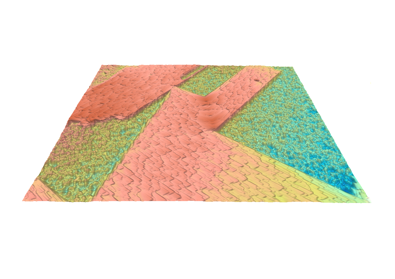
SEM Topography
Hardware
Turnkey add-on system for any SEM
- Backscattered electron detector
- Multi-channel video processor
- Scan generator and image acquisition
- Live surface topography software
Multi-segment solid-stage backscattered electron detectors
- Monolithic or hybrid quadrant sensors for high resolution
- Optional light blind sensors for high temperature/in situ SEM
- In situ pre-amplification for minimum noise and maximum speed
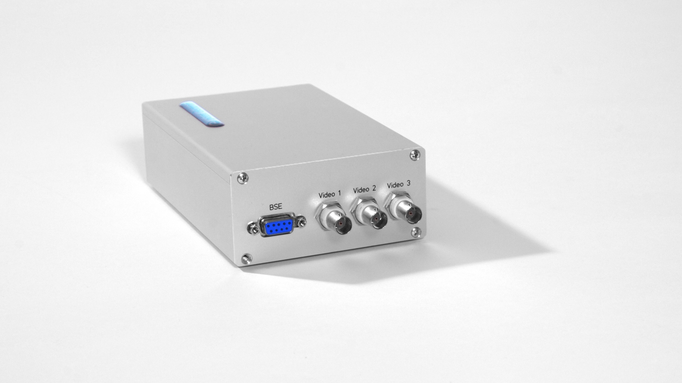
Video processor for simultaneous data acquisition
- Channel independent brightness and contrast controls
- Hardware mixed output for simultaneous acquisition with SE, BSE, EBIC, CL, or EDS
- USB controlled and fully integrated with the acquisition software
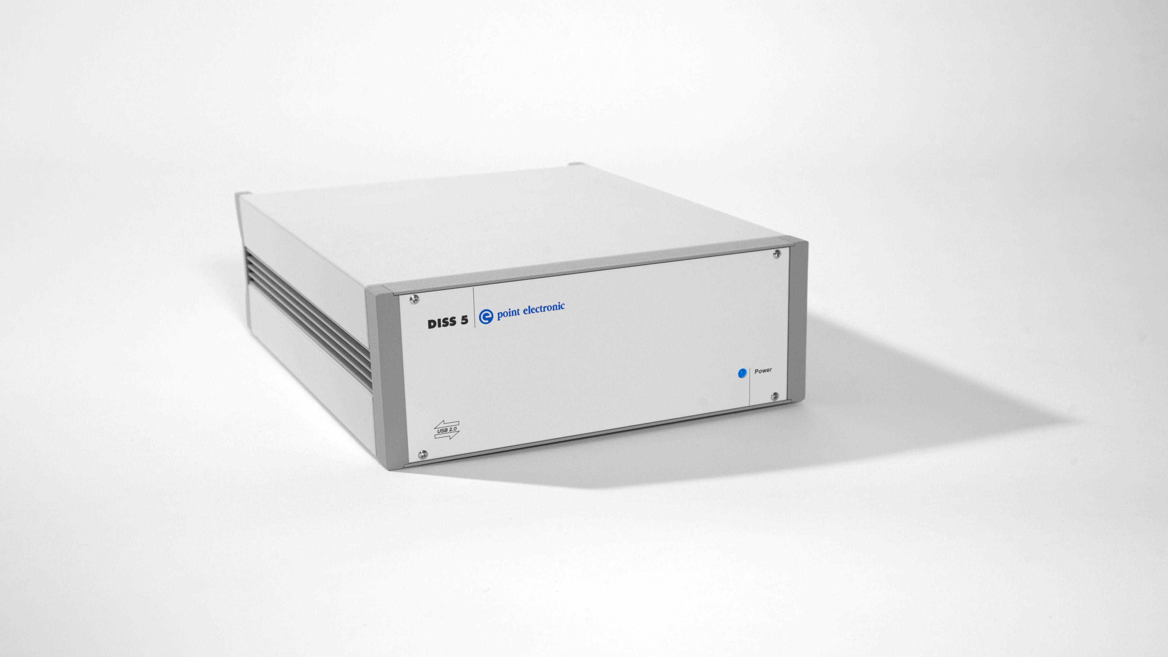
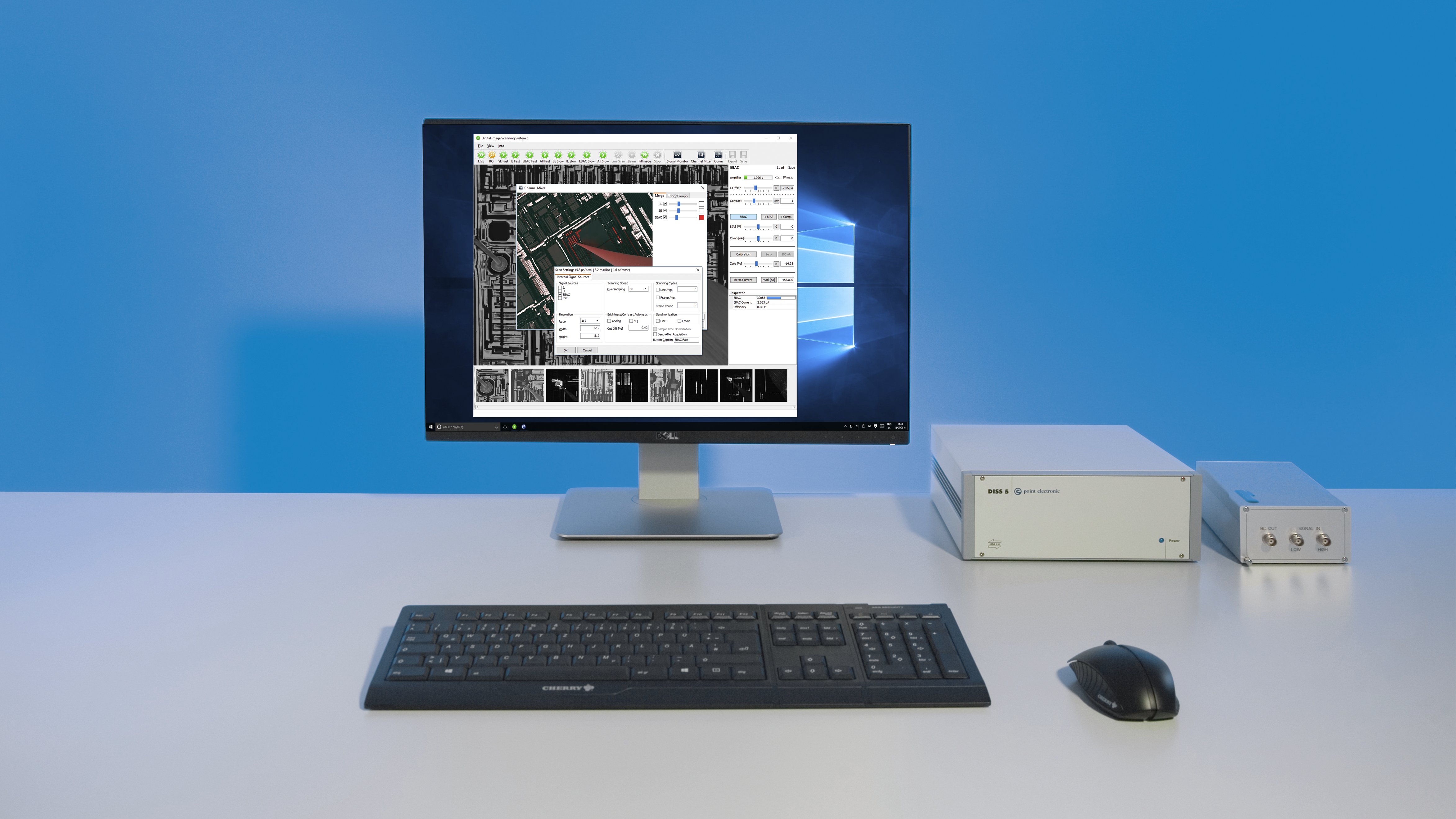
The most powerful and versatile SEM scanning system – DISS5
- Integrated scan generator and image acquisition
- Very large image resolution, up to 16k x 16k pixels
- Very fast scanning speed, down to 200ns dwell time
- Simultaneous 4x analogue and 12x digital counter inputs
Software
Completely integrated control and acquisition
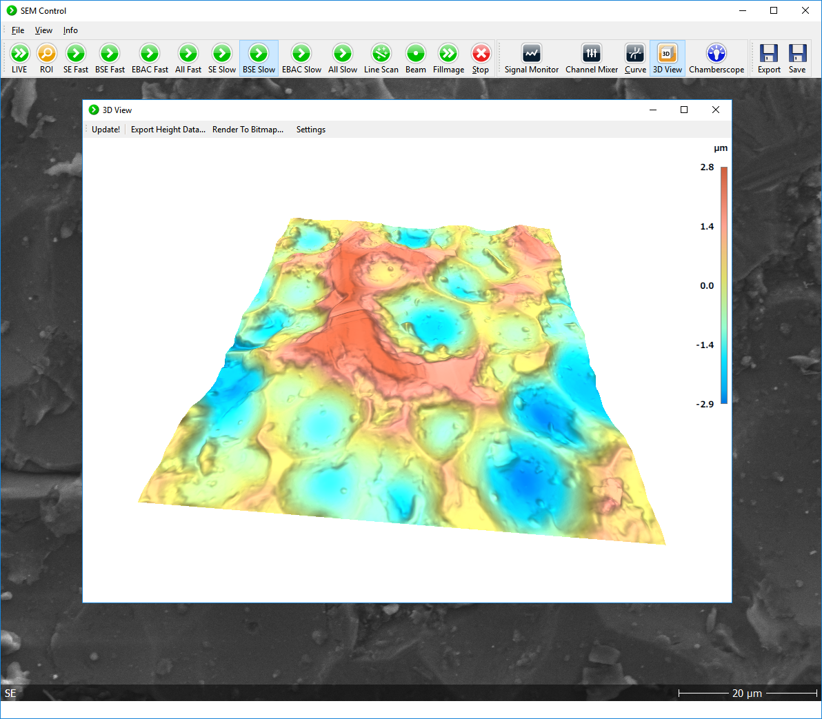
- Live surface height reconstruction from BSE signals
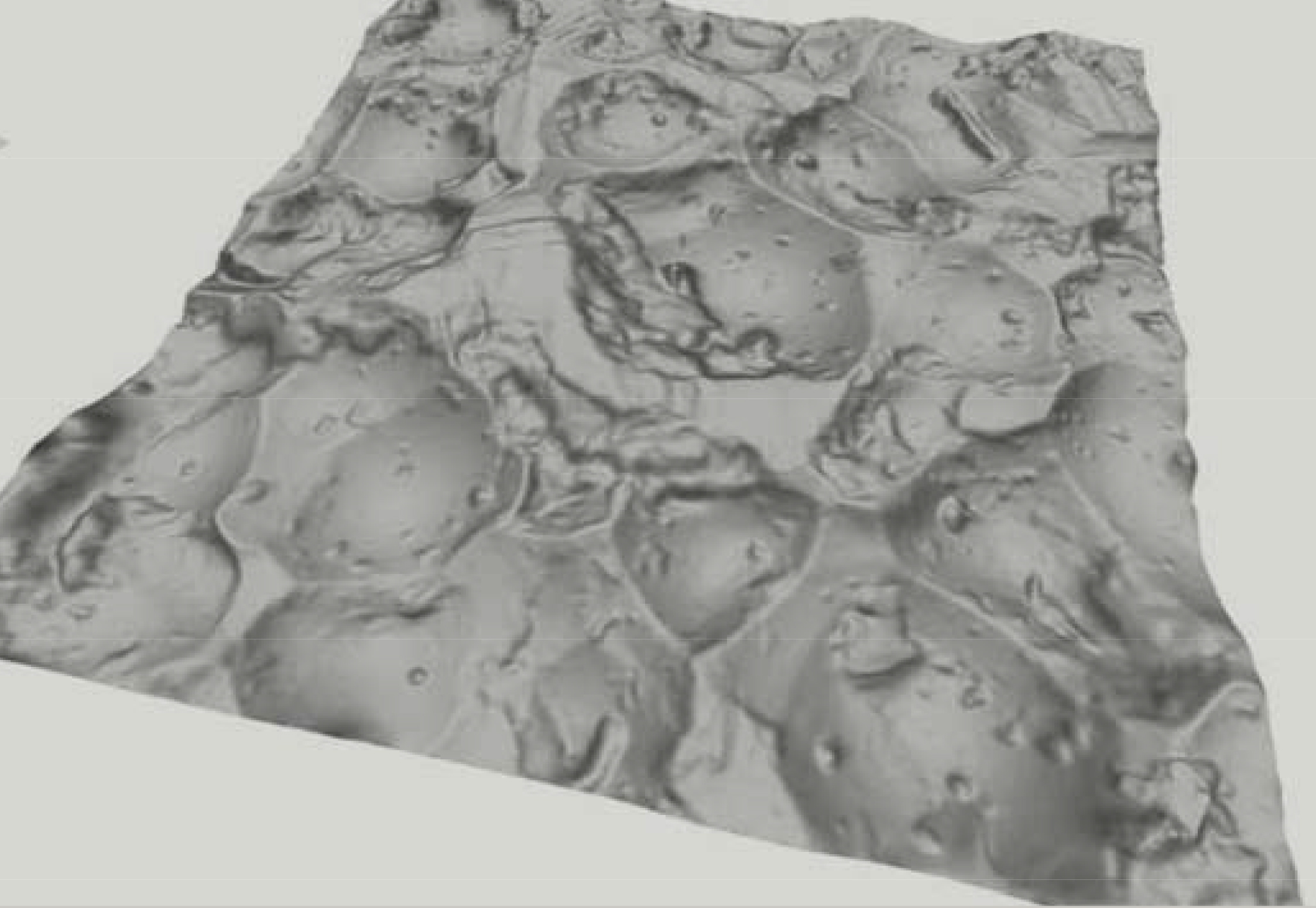
- Built-in 3D surface visualisation tool
- Configurable workflows with integrated SE and BSE scan profiles
Reconstructed height is purely topographic and quantitative
- Surface normals are calculated from 4x BSE gradients at each beam position
- Complete surface topography is assembled from all surface normals
- Height resolution depends primarily on beam/sample interaction volume
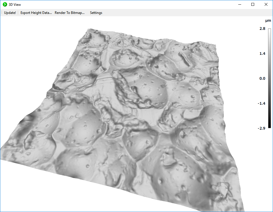
Live height visualisation tool
- Pan, rotate, tilt, zoom and scale Z
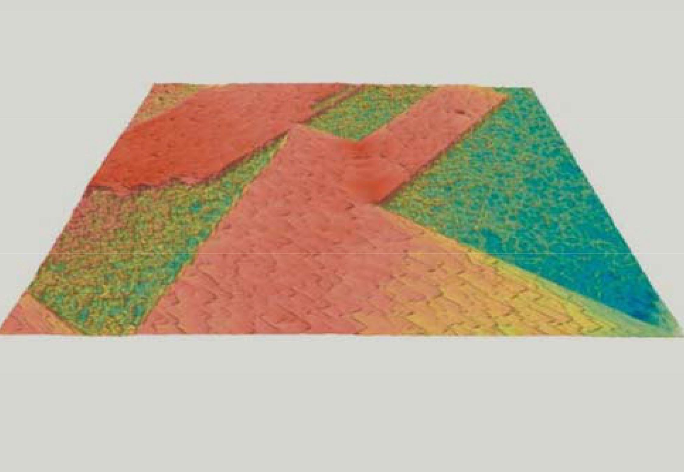
- Enhance with shadows and custom colour coding
- Export to standard binary AL3D and plain text SDF formats
One step calibration software with dedicated SEM standard – m2c
- Calibration of scale in all three spatial directions
- Orthogonality of all axes (shearing)
- Analysis of nonlinear deviations
Measure surface topography with SEM
- Use conventional segmented BSE signals
- Get immediate feedback with automated topographic reconstruction
- Save topographic data in standard surface file formats
Distinguish between sample composition and surface topography
- Resolve ambiguities in image interpretation
- Reach a wider audience with 3D models, visualisation and printing
- Measure 3D distances, volumes, steps, roughness, texture…
Monitor FIB milling and GIS deposition
- Improve yield of TEM and atom probe sample preparation
- Verify thickness of GIS deposited layers
- Monitor FIB milling depths and roughness
Visualise complex surfaces with ease
- Add texture from SE, EDS or EBSD maps
- Manipulate automatic colour textures
- Extract 3D screenshots for high-impact visualisation
Calibrate and measure height from offline BSE data
- Calibrate measurements with dedicated calibration sample
- Measure 3D positions, distances and angles
- Measure height and texture for visualisation and analysis
Continue live topography workflow with off-line analysis
- Import data into full feature analysis software
- Measure surface roughness and analyse texture
- Analyse morphology, grain and particle distributions
Backscattered (BSE) detector
- Sensor geometry: 4x quadrant diodes (monolithic sensor), or 4x rectangular diodes (hybrid sensor)
- Sensor min. energy: 1keV for detector grade Si diodes, or 5keV for standard Si diodes
- Sensor mount: retractable arm, or pole piece mount
- Sensor height: 1.2 mm
- Pre-amplifier mount: in situ
- Pre-amplifier gain: 10^7 V/A fixed
- Pre-amplifier size: 22 x 18 mm
Video processor module
- Hardware interface: USB 2.0
- BSE signal inputs: 4x max.
- SE and AUX signal inputs: 3x max.
- Simmultaneous output signals: 4x max.
- Mixed BSE output: 1x conventional compo.
- Gain: 1 … 100x
- Output offset: 1.25V max
- Brightness and Contrast control: integrated software control
- Signal inversion: Yes
- Low-pass filter: 8-levels
Topography calibration sample (m2c)
- GIS deposited calibration structures: 3x multi-level pyramidal, 1x spherical element
- FIB milled recognition structure: nanomarkers
- Total area: 80 x 80 μm, or 40 x 40 μm
- Nanomarker geometry: 800 nm, or 600 nm
- Pyramidal element geometry: 90 x 90 x 3 µm, or 45 x 45 x 1.8 µm
- Spherical element geometry: 10 x 10 x 1 µm
- Reference data: CD/ROM
SEM topography acquisition module (DISS 5 Topography)
- Hardware interface: USB 2.0
- Simultaneous inputs (i.e. SE, BSE): 4x, 12-bit
- Mapping signals (i.e. EDS): 12x, 12-bit
- Scanning interface: pre-configured for SEM and analytical add-ons
- Synchronization interface: pixel, line, frame
- Scan size: 16,384 x 16,384 pixels max.
- Pixel dwell time: 200 ns … 6 milliseconds
- Pixel over-sampling: 32,000x max.
- Line averaging: 50x max.
- Frame averaging: 256x max.
- Synchronization: mains power
- ROI scan: yes
PC/Laptop, Display
- PC/Laptop: Intel Core i3, 2x USB 2.0 minimum
- Display: 1,280 x 1,024 resolution minimum, 2x displays recommended
- Operating systems: Windows 10 … Windows XP
- Network connection: recommended
Acquisition software (DISS 5 Topography)
- BSE detector control: complete software integration
- SEM mag, kV information: automated SEM communication
- SEM topography reconstruction: live automatic shape-from-shade (m2c)
- SEM topography display: 3D visualisation, mountain view
- SEM topography control: live rotation, pan, tilt, zoom, height-rescalling
- SEM topography texture: solid colour, height-coded colour LUT
- Image mixing tool: independent colour/channel, independent intensity/channel, topographic and compositional profiles
- Scan profile templates: live display, region of interest (ROI), slow scan, area mapping, line scan, video recording (AVI)
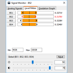
- Signal monitor: analogue signals profiles, level meter graphs, gradation graph tool
- Software API: yes
- Image caption overlay: yes
- Default BSE image file formats: 8 and 16-bit multi-page TIFF
- Default topography file formats: AL3D, SDF
- Export image file formats: BMP, JPEG, PNG, GIF
- Export topography file formats: BMP
- Operating system: Windows 10 … Windows XP
- Context sensitive help: English, German
Analysis software (microCal and micro-Shape from m2c)
- XYZ calibration: one step calibration with dedicated calibration structure
- Data extraction: point and line profiles
- 3D measurements: distances, angles
- Save file formats: BCR, BCRF, AL3D, NMM, ASC
Export file formats: PLY, TIF, BMP, JPG, PNG, GIF, TXT, DAT - Operating system: Windows 10 … Windows XP
- Context sensitive help: English, German
Parts and Cables
- BSE sensor: optional 1x
- BSE pre-amplifier: optional 1x
- BSE insertion/retraction mechanism: optional 1x
- BSE in situ cable: optional 1x
- BSE vacuum electrical feedthrough: optional 1x
- BSE ex situ cable: standard 1x
- Topography calibration sample: standard 1x
- Video processor module: standard 1x
- Video processor ground strap: standard 1x
- SEM mixed control & acquisition cable: standard 1x
- SEM acquisition module: standard 1x
- USB cables: standard 2x
- USB memory stick: standard 1x
- PC, keyboard, mouse: optional 1x
- Displays: optional 2x
Software packages
- Video Processor USB driver: Video Processor
- SEM acquisition USB driver: Digital Image Scanning System 5
- Shape-from-shadow library: m2cMicroShape (m2c)
- Acquisition software: DISS 5
- Analysis software: microShape (m2c)
- Calibration software: microCal (m2c)
Weight and Dimensions
- Video processor dimensions: 19 x 10.5 x 5 cm
- Video processor weight: 1.1 kg
- DISS5 module dimensions: 23.5 x 8.7 x 29.5 cm
- DISS5 module weight: 3.4 kg
- Shipping dimensions: typ. 36 x 32 x 56 cm
- Shipping weight: typ. 7.5 kg
Site requirements
- Power: 2x mains 110/220 VAC single phase 50-60 Hz, on the same earth as the microscope
- SEM chamber: 1x flange prepared for muti-pin vacuum electrical feedtrough to pre-amplifier
- SEM earth: 1x ground strap
- SEM interface: 1x external analogue interface for XY scan control and SEM signals
- Space: video processor module must be placed in close proximity of the SEM chamber, power supply and DISS5 modules may be placed on the SEM bench


EBAC Aquisition System
Hardware
The system is fully integrated and software controlled
- Image acquisition and amplification control are integrated
- All amplification and acquisition parameters are software controlled
- Acquisition system is compatible with all nanoprobers and SEM, including FIB/SEMs
Lowest noise EBAC/RCI amplification with in situ pre-amplifier
- In situ preamplification is optimised and fixed at 10^7 V/A
- Second stage amplification optimizes signal for image acquisition
- Signal is digitized at 12.bit to minimise quantification noise
The most powerful and versatile SEM scanning system – DISS5
- Integrated scan generator and image acquisition
- Very large image resolution, up to 16k x 16k pixels
- Very fast scanning speed, down to 200ns dwell time
- Simultaneous 4x analogue and 12x digital counter inputs
Software
The system is software controlled for efficiency and ease-of-use
- Image scanning and amplifier control are integrated into one app
- All amplification and acquisition parameters are accessible to end user
- Signal is automatically quantified and displayed in current units (µA, nA, pA)
Simultaneous signals are mixed live for localisation
- Up to 4x simultaneous signals
- 12-bit digitization with signal integration (oversampling)
- Live colour mixing tool for visualisation
Current-voltage (IV) tool is integrated for assisted touchdown
- Voltage output maximum range is -10…+10 V
- Gain selection for current measurements is automatic
- IV data may be exported in standard formats
Configurable scan profiles enable custom workflows for efficient use
- Fast EBAC scan profile for alignment and navigation
- Simultaneous SE/EBAC scan profile for localization
- High resolution EBAC scan profile for mapping and analysis
Live signal monitor assists image acquisition and calibration
- Live line scan signals are displayed for optimisation
- Multiple live signals are displayed simultaneously
- The gradation graph improves display of complex shadows
Advanced modes include biasing, beam current and calibration
- Bias is DC and includes current compensation
- Voltage bias is also included for voltage contrast
- All amplifier settings can be saved and reloaded
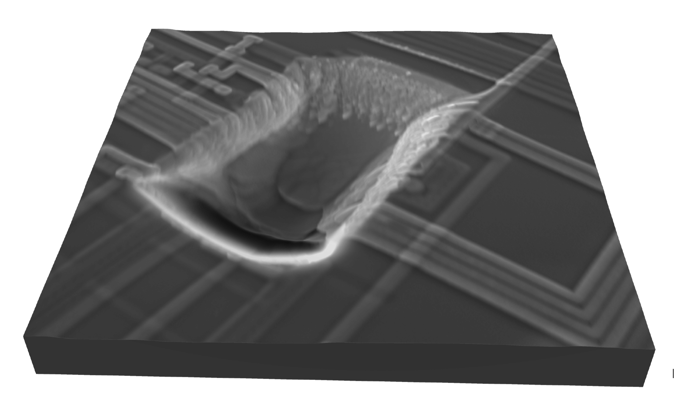
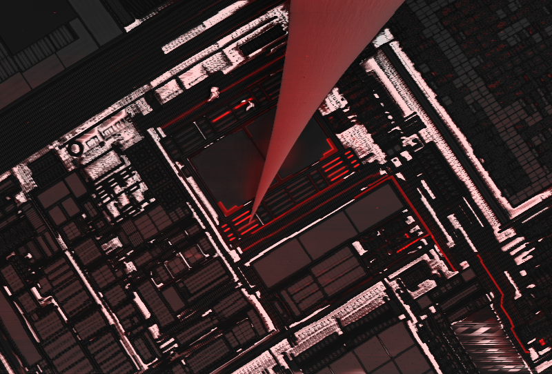
Characterize interconnects with the highest resolution
- Reveal electrical integrity of nets with sub-micron resolution
- Diagnose contamination, metal patterning defects, resistive interconnectors
- Directly isolate defects to exact layer and die location
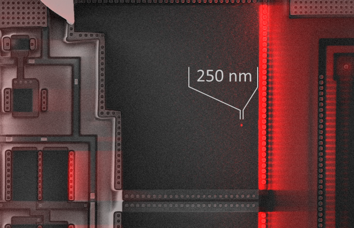
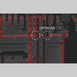
Find exact location of any open, resistive or shorting defect
- Localize metal line cuts caused by cracking, corrosion, electro-migration or foreign particles
- Identify resistive opens caused by interface contamination at vias
- Pinpoint location for direct TEM lamella FIB preparation
Verify device operation models with built-in biasing for voltage contrast
- Image bias/voltage contrast in delayered devices
- Monitor operation of devices under bias
- Compare imaged behaviour with device design
Localize defects in thin dielectric layers
- Visualize weaknesses in GOX or COX before breakdown
- Pinpoint oxide shorts caused by ESD or EOS with sub-micron resolution
- Preserve the original defect with nW power dissipation during analysis
Access failures invisible in voltage contrast
- Find low resistances that allow charge tunnelling through interconnects
- Investigate structures in contact with the silicon substrate
- Characterize large metal structures
EBAC/RCI pre-amplifier module
- Gain: 10^7 V/A fixed
- Mount: in situ, on the SEM sample stage
- Board type: standard PCB
EBAC/RCI amplifier module
- Hardware interface: USB2.0
- Input offset/brightness: -1 … 1 µA, 16-bit
- Signal gain/contrast: 0.1 … 100x, 16-bit
- Signal inversion: yes
- Bias voltage: -10 … 10 V, 16-bit
- Current compensation: -10 … 10 µA, 16-bit
- Low-pass filter: 8-levels
- Zero-balanced/calibrated pre-amplifier: manual
- Beam blank output: TTL
EBAC/RCI and SEM acquisition module (DISS5 EBAC/RCI)
- Hardware interface: USB 2.0
- Simultaneous inputs (i.e. SE, EBIC): 4x, 12-bit
- Mapping signals (i.e. EDS): 12x, 12-bit
- Scanning interface: pre-configured for SEM and analytical add-ons
- Synchronization interface: pixel, line, frame
- Scan size: 16,384 x 16,384 pixels max.
- Pixel dwell time: 200 ns … 6 milliseconds
- Pixel over-sampling: 32,000x max.
- Line averaging: 50x max.
- Frame averaging: 256x max.
- Synchronization: mains power
- ROI scan: yes
Electrical sample holder
- Contacting probes: 2x
- Contacting geometries: 1x top and 1x bottom, or 2x top
- Imaging geometry: plan-view and cross-section
- Faraday cage: 2x
- Maximum sample size: typ. 25 x 50 mm
- Holder height: 20 mm
PC/Laptop and Display
- PC/Laptop: Intel Core i3, 1x USB 2.0 minimum
- Display: 1,280 x 1,024 resolution minimum, 2x displays recommended
- Operating systems: Windows 10 … Windows XP
- Network connection: recommended
Acquisition software (DISS5 EBAC/RCI)
- EBAC/RCI control: complete software integration
- EBAC/RCI operation modes: standard, biased, calibration
- EBAC/RCI configuration: complete profile save/load
- EBAC/RCI inspector: quantified live current readings
- Current voltage (IV) tool: configurable voltage range, number of points, settle time
- SEM mag, kV information: automated SEM communication
- Brightness & Contrast controls: manual/automatic, channel independent control
- Image mixing tool: independent colour/channel, independent intensity/channel, topographic and compositional profiles
- Scan profile templates: live display, region of interest (ROI), slow scan, area mapping, line scan, video recording (AVI)
- Signal monitor: analogue signals profiles, level meter graphs, gradation graph tool
- Software API: yes
- Image caption overlay: yes
- Default file formats: 8 and 16-bit multi-page TIFF
- Export file formats: BMP, JPEG, PNG, GIF
- Operating system: Windows 10 … Windows XP
- Context sensitive help: English, German
Analysis software (DIPS5 EBAC/RCI)
- Current quantification: automated data management
- Magnification/scale information: automated scale management
- Image caption overlay: configurable live and image export overlays
- Image information display: all relevant acquisition parameters
- Image mix (SE, EBIC, etc.): configurable colour assignment
- Editable LUT (look up table): false colour, GGR file format
- Pseudo-surface view: pan/zoom/rotation/tilt controls, emission/ambient/diffuse lighting, original texture/false colouring
- Distance, area measurements: live display
- Data extraction: multiple lines/points, simultaneous signal extraction, line/signal diagrams, optional smoothing, absolute/relative scaling
- Operating system: Windows 10 … Windows XP
- Context sensitive help: English, German
Parts and Cables
- EBAC/RCI in situ pre-amplifier module: standard 1x
- EBAC/RCI sample holder: standard 1x
- EBAC/RCI in situ pre-amplifier cable: standard 1x
- EBAC/RCI electrical vacuum feedthrough: standard 1x
- EBAC/RCI ex situ pre-amplifier cable: standard 1x
- EBAC/RCI ex situ amplifier module: standard 1x
- EBAC/RCI ground strap: standard 1x
- EBAC/RCI power supply: standard 1x
- EBAC/RCI and SEM mixed control & acquisition cable: standard 1x
- EBAC/RCI and SEM acquisition module (DISS5 EBAC/RCI): standard 1x
- USB cables: standard 2x
- USB memory stick: standard 1x
- PC, keyboard, mouse: optional 1x
- Displays: optional 1x
Software Packages
- EBAC/RCI acquisition driver: EBAC/RCI Amplifier
- SEM acquisition driver: Digital Image Scanning System 5
- Acquisition software: DISS 5
- Analysis software: DIPS 5
Weight and Dimensions
- EBAC/RCI amp. module dimensions: typ. 25 x 10.5 x 6 cm
- EBAC/RCI amp. module weight: typ. 1.2 kg
- EBAC/RCI power supply dimensions: typ. 17.5 x 11 x 5.5 cm
- EBAC/RCI power supply weight: typ. 1.2 kg
- DISS5 module dimensions: 23.5 x 8.7 x 29.5 cm
- DISS5 module weight: 3.4 kg
- Shipping dimensions: typ. 36 x 32 x 56 cm
- Shipping weight: typ. 7.5 kg
Site requirements
- Power: 2x mains 110/220 VAC single phase 50-60 Hz, on the same earth as the microscope
- SEM chamber: 1x flange prepared for muti-pin vacuum electrical feedtrough to pre-amplifier
- SEM earth: 1x ground strap
- SEM interface: 1x external analogue interface for XY scan control and SEM signals
- Space: EBAC/RCI amplifier module must be placed in close proximity of the SEM chamber, EBAC/RCI power supply and DISS5 modules may be placed on the SEM bench

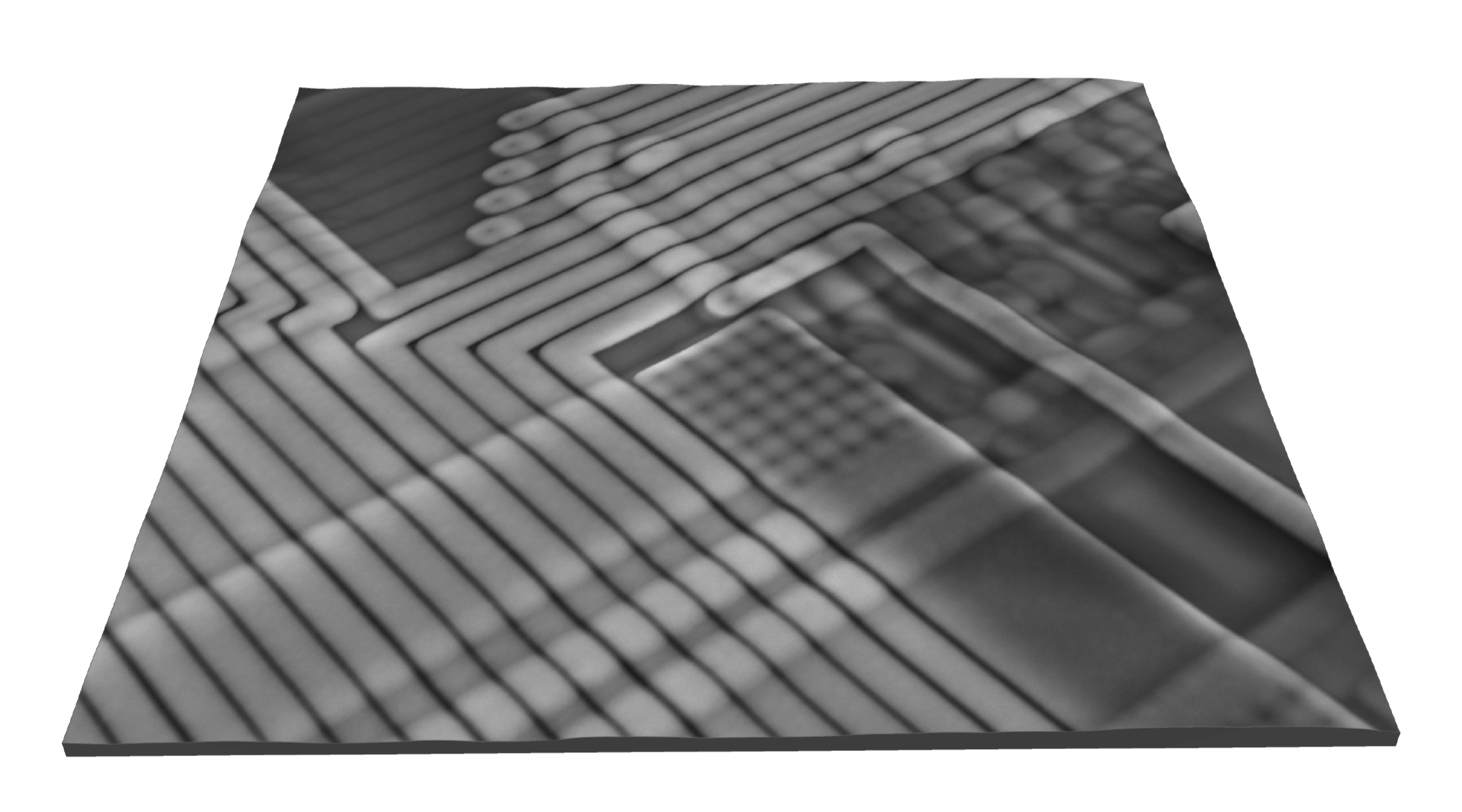
EBIC acquisition

Point Electronic GmbH: EBIC acquisition system
Hardware
The EBIC system is fully integrated and software controlled
- Image acquisition and EBIC module are integrated into one software
- All amplification and acquisition settings are software controlled
- EBIC signal is automatically quantified and displayed in current values (µA, nA, pA)
The most sophisticated and easy to use EBIC amplifier
- Two stage amplification for maximum gain range
- Built in -10…+10V DC bias with current compensation
- Beam current output for SEM feedback and integration
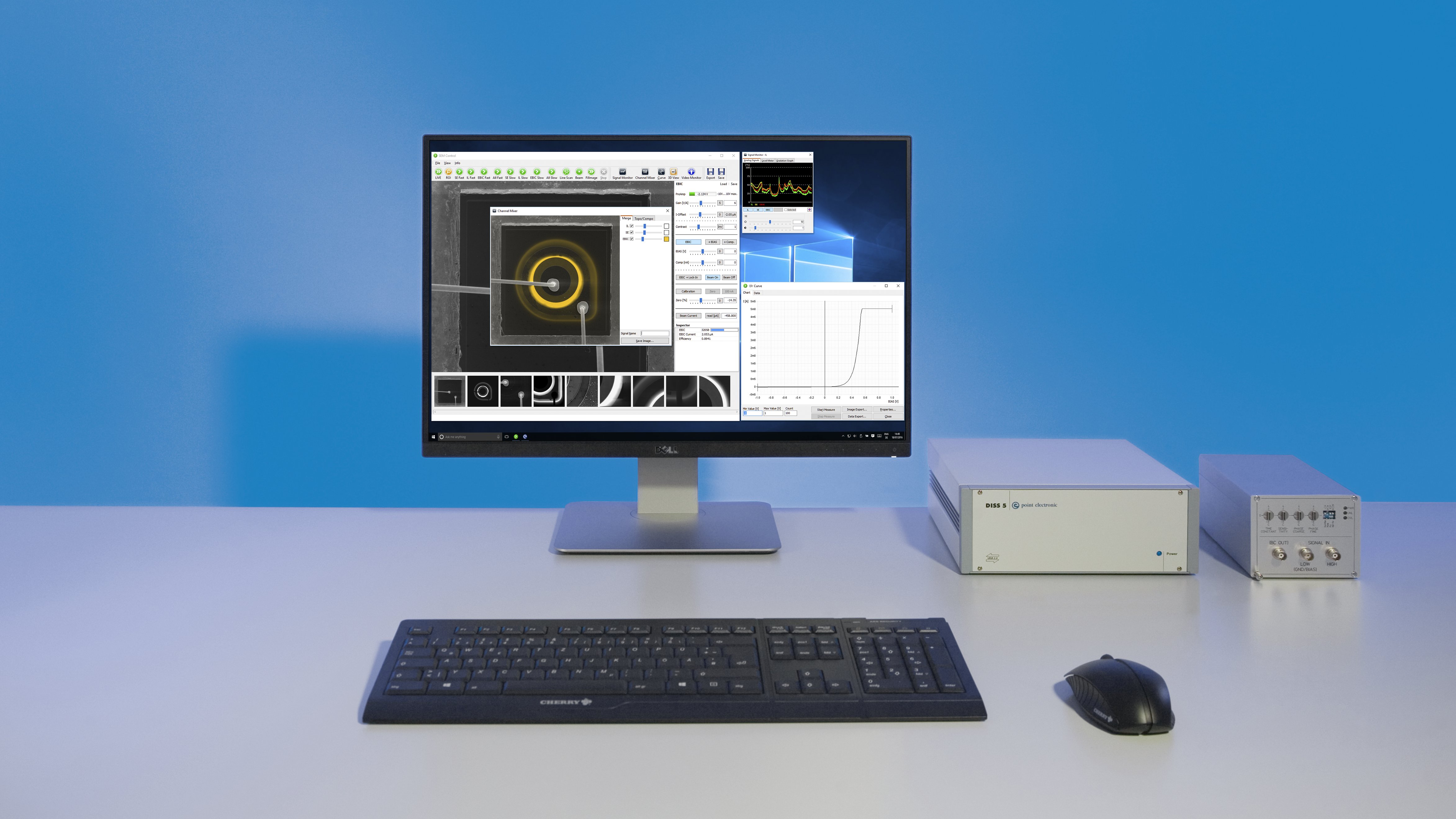
The most powerful and versatile SEM scanning system – DISS5
- Integrated scan generator and image acquisition
- Very large image resolution, up to 16k x 16k pixels
- Very fast scanning speed, down to 200ns dwell time
- Simultaneous 4x analogue and 12x digital counter inputs
Optional electrical sample holder for large area devices
- Suitable for solar cells, photovoltaics and light emitting diodes
- Flexible mount in plan-view or cross-section configuration
- Includes Faraday cups for beam current measurements
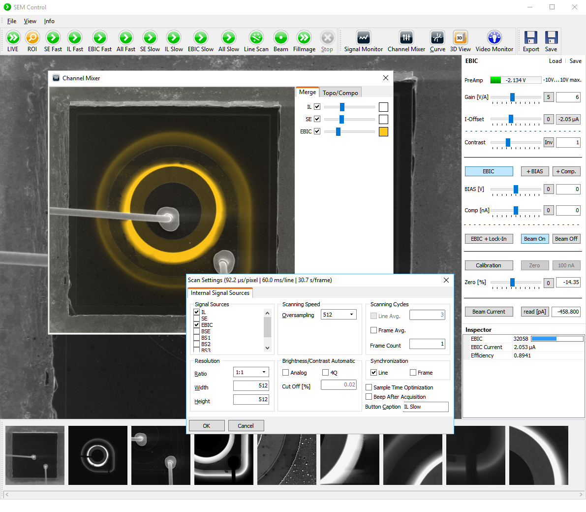
Software
Advanced controls are provided for calibration, biasing and scanning
- Flexible pre-amplifier gain from as little as 10^3 to as high as 10^10 V/A
- Further 0.1…100x gain and 100 µA compensation for optimum imaging
- Electronics are optimized for high speed, providing 0.5 MHz at 10^9 V/A
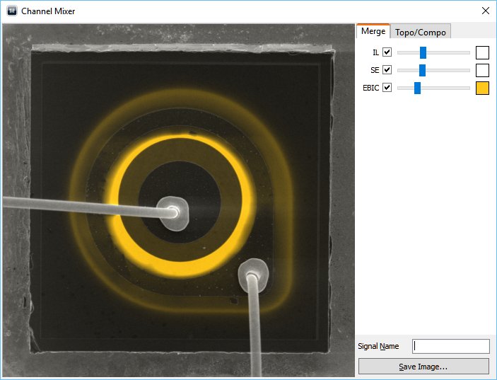
Simultaneous signals are mixed live for correlative microscopy
- Up to 4x simultaneous signals
- 12-bit digitization with signal integration (oversampling)
- Live color mixing tool for visualization
Current-voltage (IV) tool is integrated for contacts and Nano probing
- Voltage output maximum range is -10…+10 V
- Gain selection for current measurements is automatic
- IV may also be used for device characterization
Configurable scan profiles enable custom workflows for efficient use
- Fast EBIC scan profile for alignment and navigation
- High resolution EBIC scan profile for mapping and analysis
- Simultaneous SE/EBIC scan profile for localization
Live signal monitor assists image acquisition and calibration
- Live line scan signals are displayed for optimization
- Multiple live signals are displayed simultaneously
- The gradation graph improves display of complex shadows
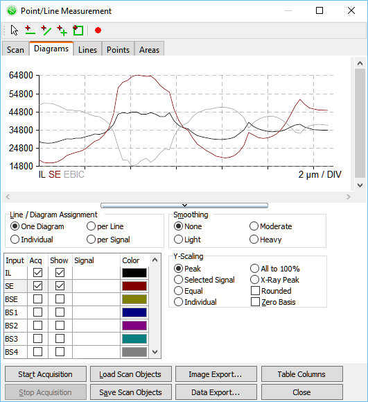
Advanced live scan tool enables advanced beam control
- Select points, lines or areas from pre-scan images
- Set number of points, step size, binning and averaging
- Generate single or multiple diagrams
- Export diagrams and/or raw data
Make the link between device characterization and materials properties
- Image electrical activity across complete devices
- Distinguish between electrically active and passive defects
- Correlate electrical activity with composition (EDS) and crystallographic structure (EBSD)
Localize electrical defects with highest resolution
- Enable sample preparation for TEM or atom probe microscopy
- Avoid alignment errors by directly imaging defects with EBIC in FIB SEM
- Use live EBIC imaging to stop milling during sample preparation
Map junctions and defects over large areas
- Identify all electrically active defects
- Map active areas of junctions and electrical fields
- Validate doping profiles and areas
Export calibrated EBIC signal for analysis of materials properties
- Measure defect contrast / recombination strength
- Extract diffusion length of minority charge carriers
- Determine width of depletion regions
Verify device operation modes with built-in biasing and live overlay
- Image junctions and fields in delayered devices
- Map electrical activity in solar cells under bias
- Compare imaged behaviour with device modelling
Access third dimension with depth profiling
- Manipulate depth of EBIC signal by changing kV in SEM
- Investigate EBIC images of cross-sections in FIB-SEM
- Export EBIC depth series for 3D reconstruction
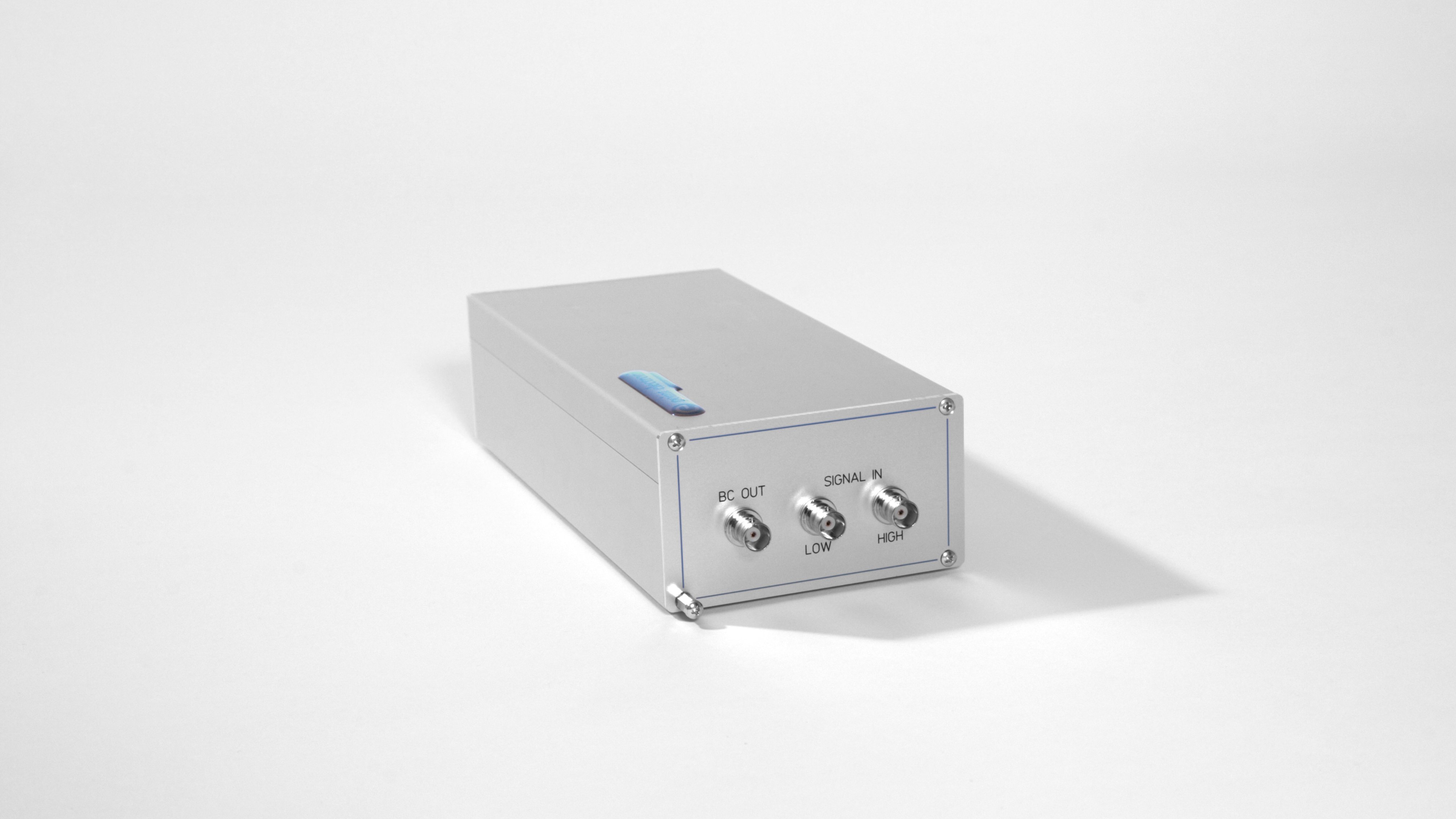
EBIC amplifier module
- Hardware interface: USB2.0
- Pre-amplifier gain: 10^3 … 10^10 V/A
- Input offset/brightness: -1 … 1 µA, 16-bit
- Signal gain/contrast: 0.1 … 100x, 16-bit
- Signal inversion: yes
- Bandwidth: 0.5 MHz at 10^9 V/A
- Bias voltage: -10 … 10 V, 16-bit
- Current compensation: -10 … 10 µA, 16-bit
- Low-pass filter: 8-levels
- Zero-balanced/calibrated pre-amplifier: manual
- Beam blank output: TTL
- Lock-in amplification (optional): yes
EBIC and SEM acquisition module (DISS5 EBIC)
- Hardware interface: USB 2.0
- Simultaneous inputs (i.e. SE, EBIC): 4x, 12-bit
- Mapping signals (i.e. EDS): 12x, 12-bit
- Scanning interface: pre-configured for SEM and analytical add-ons
- Synchronization interface: pixel, line, frame
- Scan size: 16,384 x 16,384 pixels max.
- Pixel dwell time: 200 ns … 6 milliseconds
- Pixel over-sampling: 32,000x max.
- Line averaging: 50x max.
- Frame averaging: 256x max.
- Synchronization: mains power
- ROI scan: yes
Electrical sample holder
- Contacting probes: 2x
- Contacting geometries: 1x top and 1x bottom, or 2x top
- Imaging geometry: plan-view and cross-section
- Faraday cage: 2x
- Maximum sample size: typ. 25 x 50 mm
- Holder height: 20 mm
PC/Laptop and Display
- PC/Laptop: Intel Core i3, 1x USB 2.0 minimum
- Display: 1,280 x 1,024 resolution minimum, 2x displays recommended
- Operating systems: Windows 10 … Windows XP
- Network connection: recommended
Acquisition software (DISS5 EBIC)
- EBIC control: complete software integration of EBIC hardware
- EBIC operation modes: standard, biased, calibration, external beam measurement
- EBIC configuration: complete profile save/load
- EBIC inspector: quantified live current readings
- Current voltage (IV) tool: configurable voltage range, number of points, settle time
- SEM mag, kV information: automated SEM communication
- Brightness & Contrast controls: manual/automatic, channel independent control
- Image mixing tool: independent colour/channel, independent intensity/channel, topographic and compositional profiles
- Scan profile templates: live display, region of interest (ROI), slow scan, area mapping, line scan, video recording (AVI)
- Signal monitor: analogue signals profiles, level meter graphs, gradation graph tool
- Software API: yes
- Image caption overlay: yes
- Default file formats: 8 and 16-bit multi-page TIFF
- Export file formats: BMP, JPEG, PNG, GIF
- Operating system: Windows 10 … Windows XP
- Context sensitive help: English, German
Analysis software (DIPS5)
- Current quantification: automated data management
- Magnification/scale information: automated scale management
- Image caption overlay: configurable live and image export overlays
- Image information display: all relevant acquisition parameters
- Image mix (SE, EBIC, etc.): configurable colour assignment
- Editable LUT (look up table): false colour, GGR file format
- Pseudo-surface view: pan/zoom/rotation/tilt controls, emission/ambient/diffuse lighting, original texture/false colouring
- Distance, area measurements: live display
- Data extraction: multiple lines/points, simultaneous signal extraction, line/signal diagrams, optional smoothing, absolute/relative scaling
- Operating system: Windows 10 … Windows XP
- Context sensitive help: English, German
Parts and Cables
- EBIC amplifier module: standard 1x
- EBIC power supply: standard 1x
- EBIC and SEM acquisition module: standard 1x
- Electrical sample holder: standard 1x
- Electrical vacuum feedtrough: standard 1x
- EBIC input coax cables: standard 2x
- SEM and EBIC mixed control & acquisition cable: standard 1x
- Ground strap: standard 1x
- USB cables: standard 1x
- USB memory stick: standard 1x
- PC, keyboard, mouse: optional 1x
- Displays: optional 2x
Software Packages
- EBIC acquisition driver: EBIC Amplifier
- SEM acquisition driver: Digital Image Scanning System 5
- Acquisition software: DISS 5
- Analysis software: DIPS 5
Weight and Dimensions
- EBIC amp. module dimensions: typ. 10.5 x 6.0 x 25.0 cm
- EBIC amp. module weight: typ. 1.1 kg
- EBIC power supply dimensions: typ. 11.0 x 5.5 x 17.0 cm
- EBIC power supply weight: typ. 1.2 kg
- DISS5 module dimensions: 23.5 x 8.7 x 29.5 cm
- DISS5 module weight: 3.4 kg
- Shipping dimensions: typ. 36 x 32 x 56 cm
- Shipping weight: typ. 7.5 kg
Site requirements
- Power: 2x mains 110/220 VAC single phase 50-60 Hz, on the same earth as the microscope
- SEM chamber: 1x flange prepared for muti-pin vacuum electrical feedtrough to device under test
- SEM earth: 1x ground strap
- SEM interface: 1x external analogue interface for XY scan control and SEM signals
- Space: EBIC module must be placed in close proximity of the SEM chamber, EBIC power supply and DISS5 modules may be placed on the SEM bench


4Q-BSE Detector
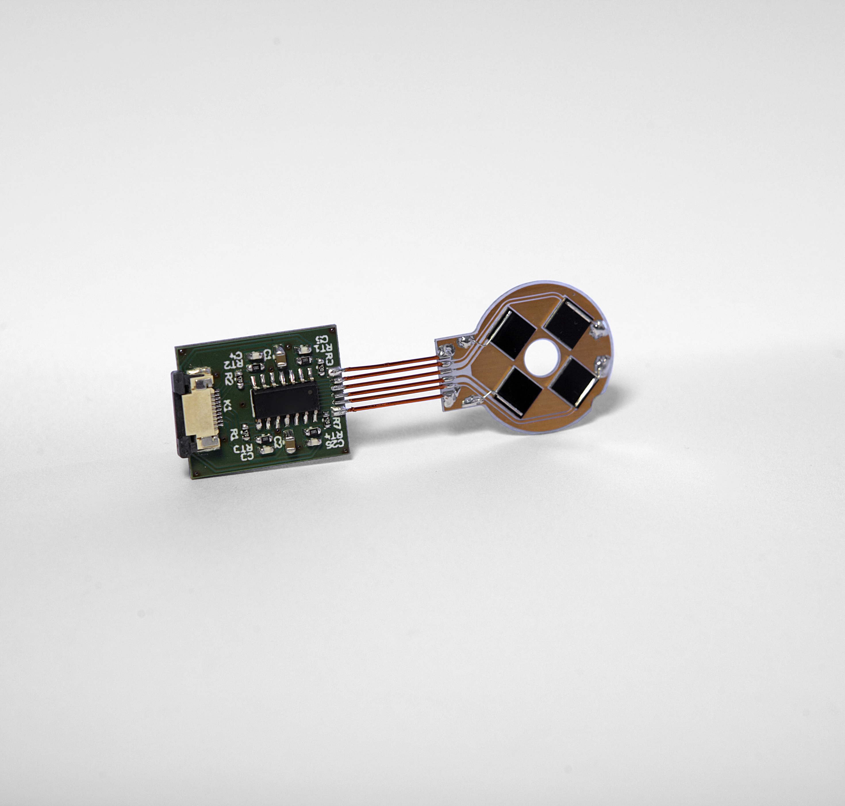
Point Electronic GmbH: 4-quadrant detector with integrated preamplifier
- 10-pin electrical feed-through
- USB2 controlled video-processor with 4 inputs for the BSE-detector, 3 inputs for SE, CL, …
- adjustment of brightness and contrast
- 8 step low-pass filter
- DISS5 software-module for video-processor control
- easy adjustment of Topo- and Compo-images in the DISS5 Channel Mixer
- simultaneous acquisition of the 4 separate BSE-signal
- optional live SEM Topography function in DISS5
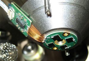
Four-quadrant solid-state backscatter electron detector provides both topography and materials contrast (composition) imaging.
Technical Details
- Integrated preamplifier, mounted below the objective-lens
- Size of the detector: Ø 25 mm, h 1.2 mm
- Size of the preamplifier: 22 × 18 mm

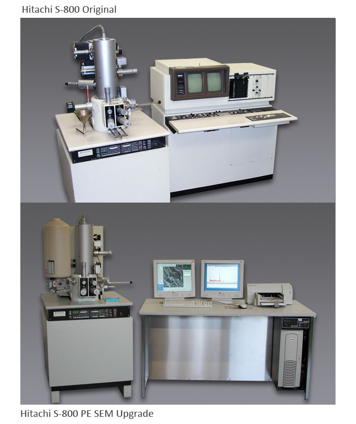
SEM Upgrades
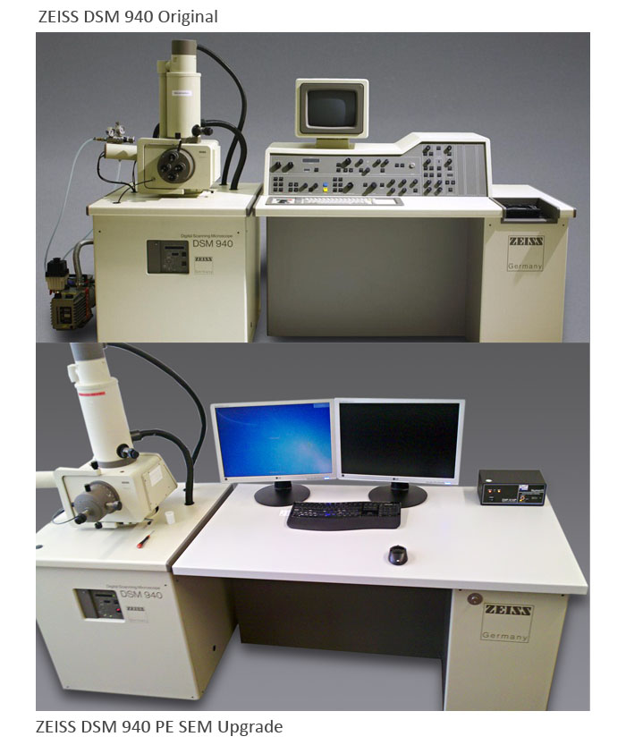
Remove the old, failing electronics from your SEM and upgrade with NEW Windows 10 compatible control systems.
We replace all of the old electronics, including the power supplies, scan system, and imaging system, and we can even replace the SE and Backscatter detectors with new hardware- keep your existing column and all the detector configuration you’ve invested so much in, but get a state-of-the-art new microscope control system from Point Electronic.
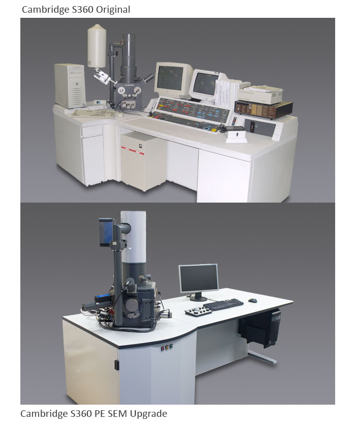
Upgrades
Remove the old, failing electronics from your SEM and upgrade with NEW Windows 10 compatible control systems.
We replace all of the old electronics, including the power supplies, scan system, and imaging system, and we can even replace the SE and Backscatter detectors with new hardware- keep your existing column and all the detector configuration you’ve invested so much in, but get a state-of-the-art new microscope control system.

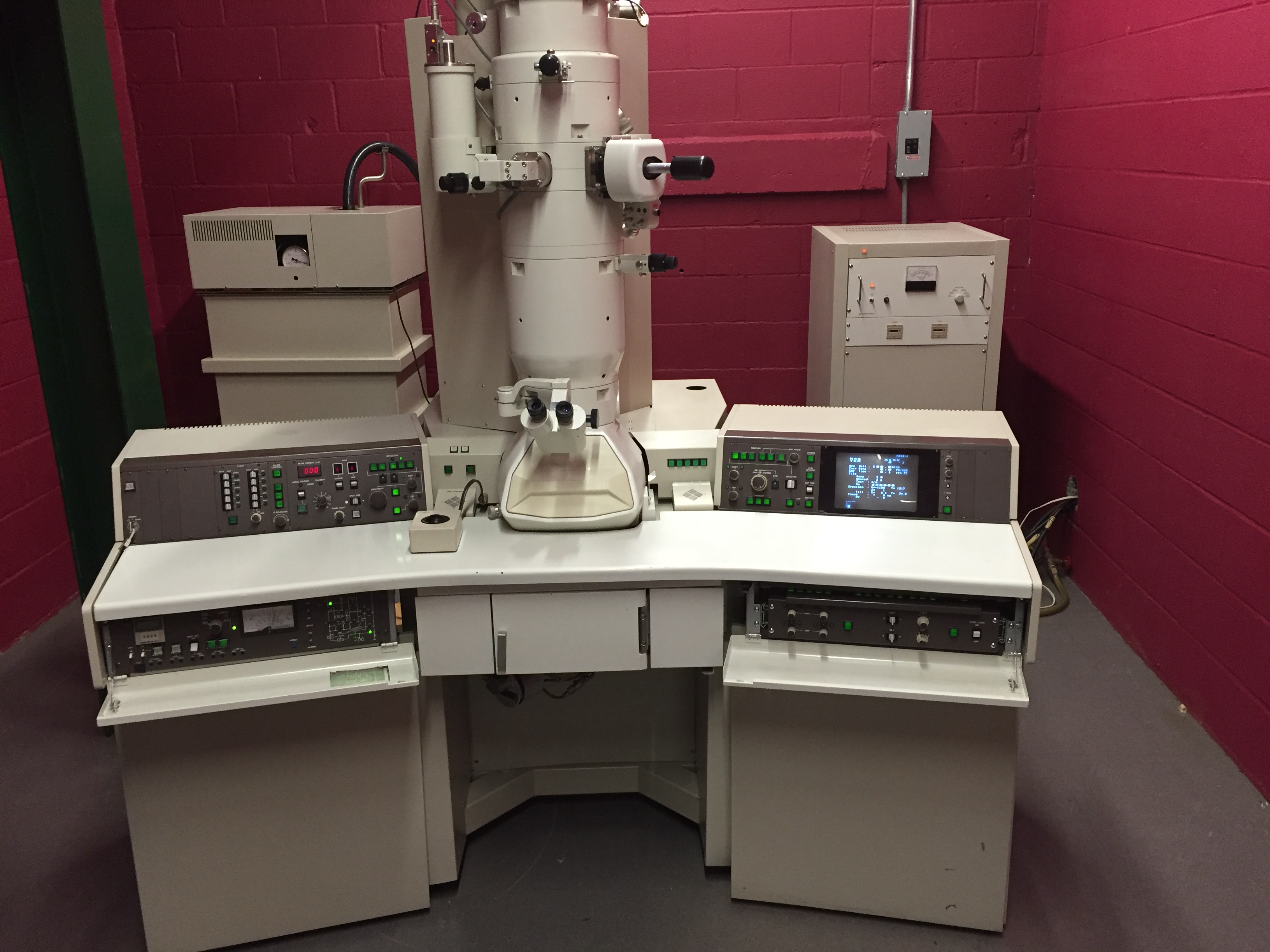
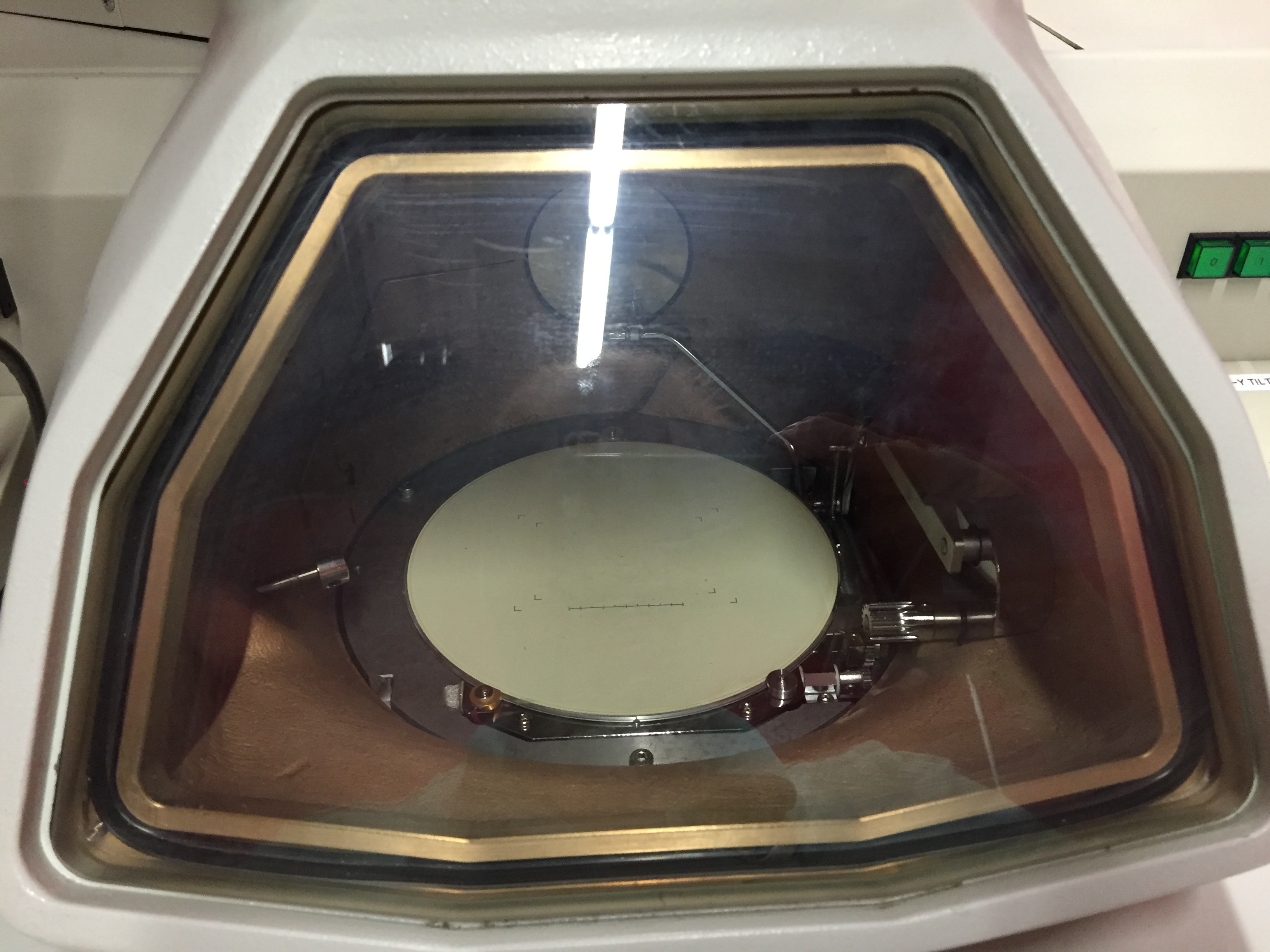
JEOL 2010
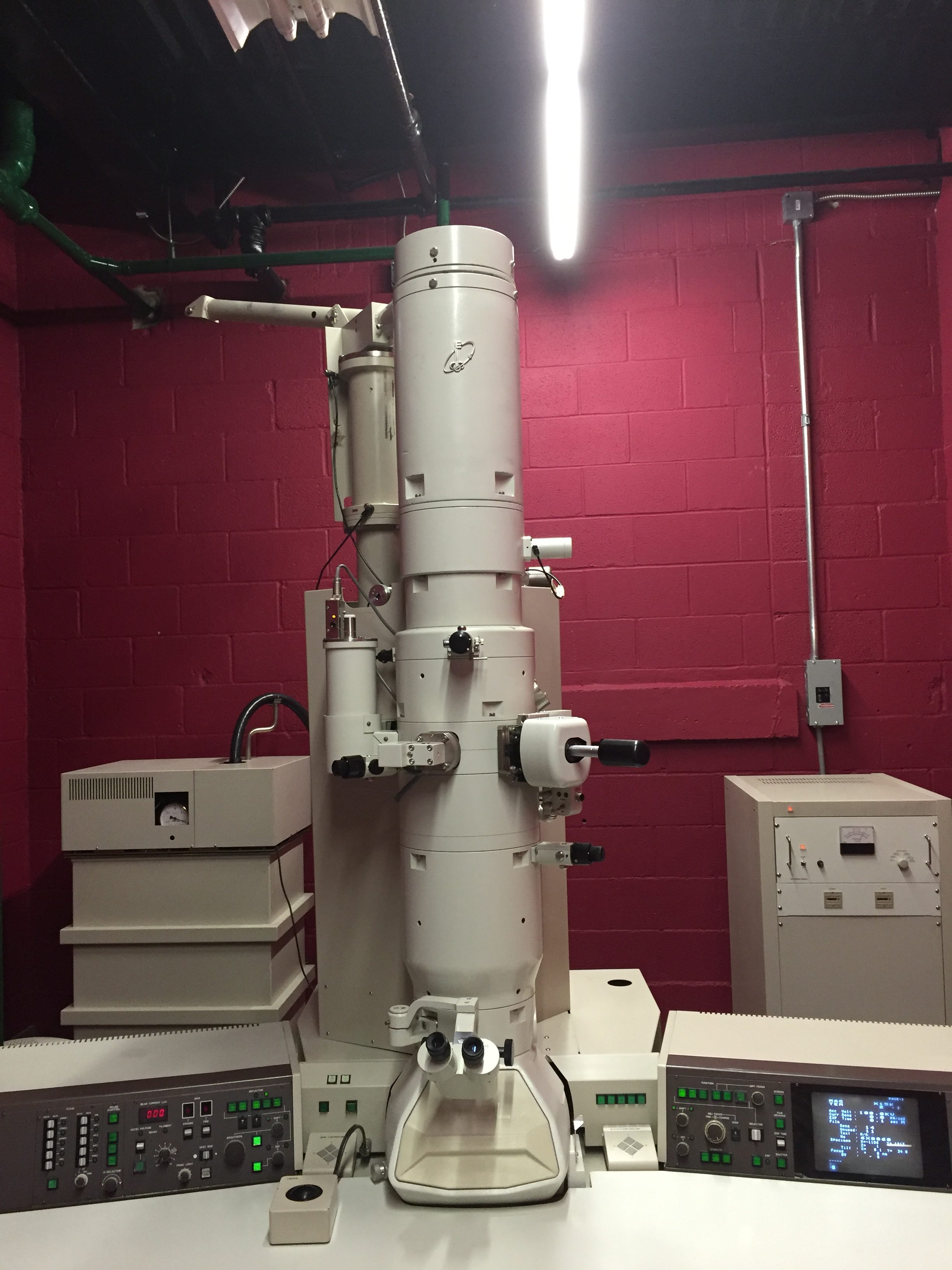
JEOL 2010 200KV TEM
Camera and EDS available
Delivery and installation available

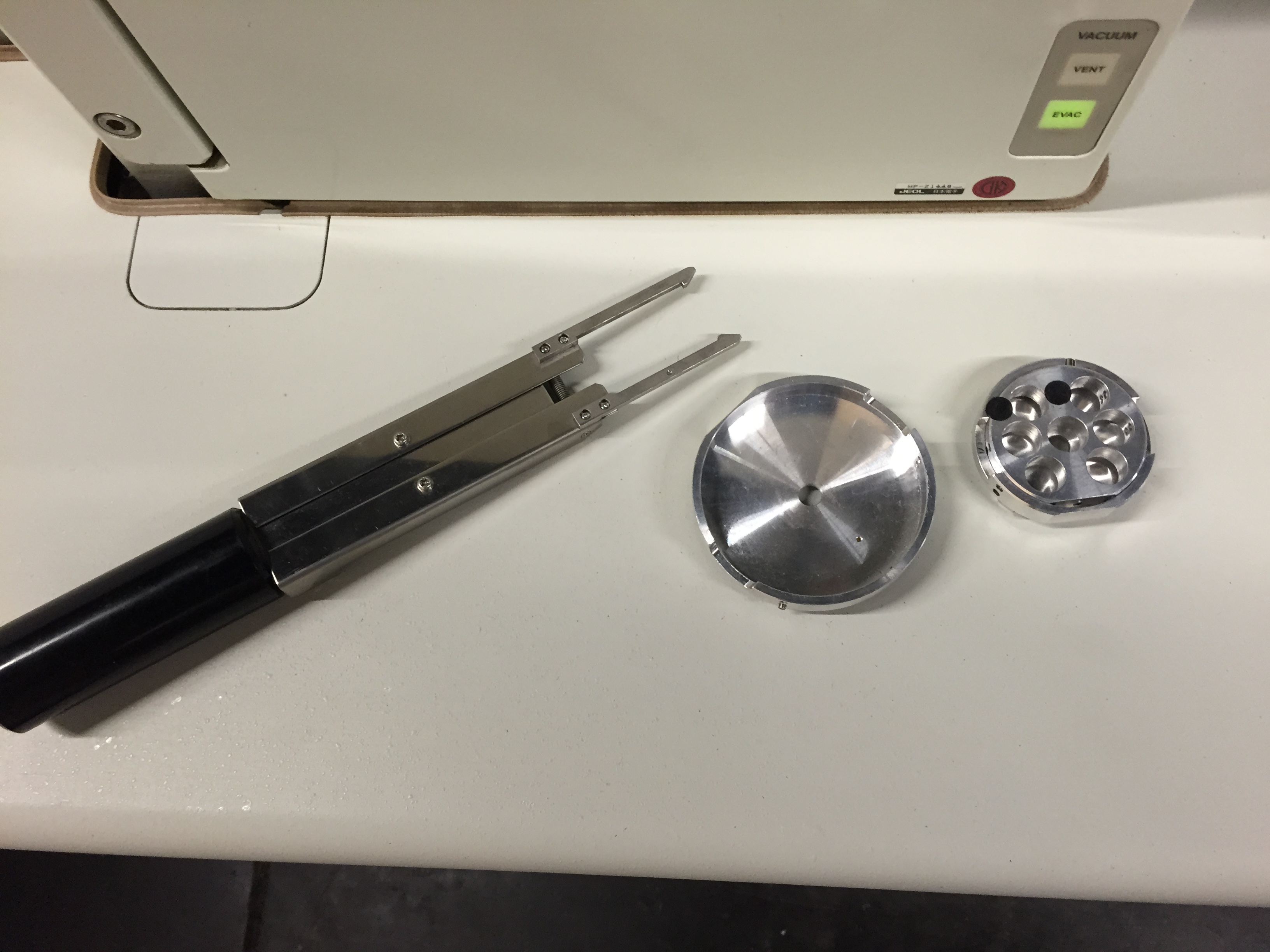
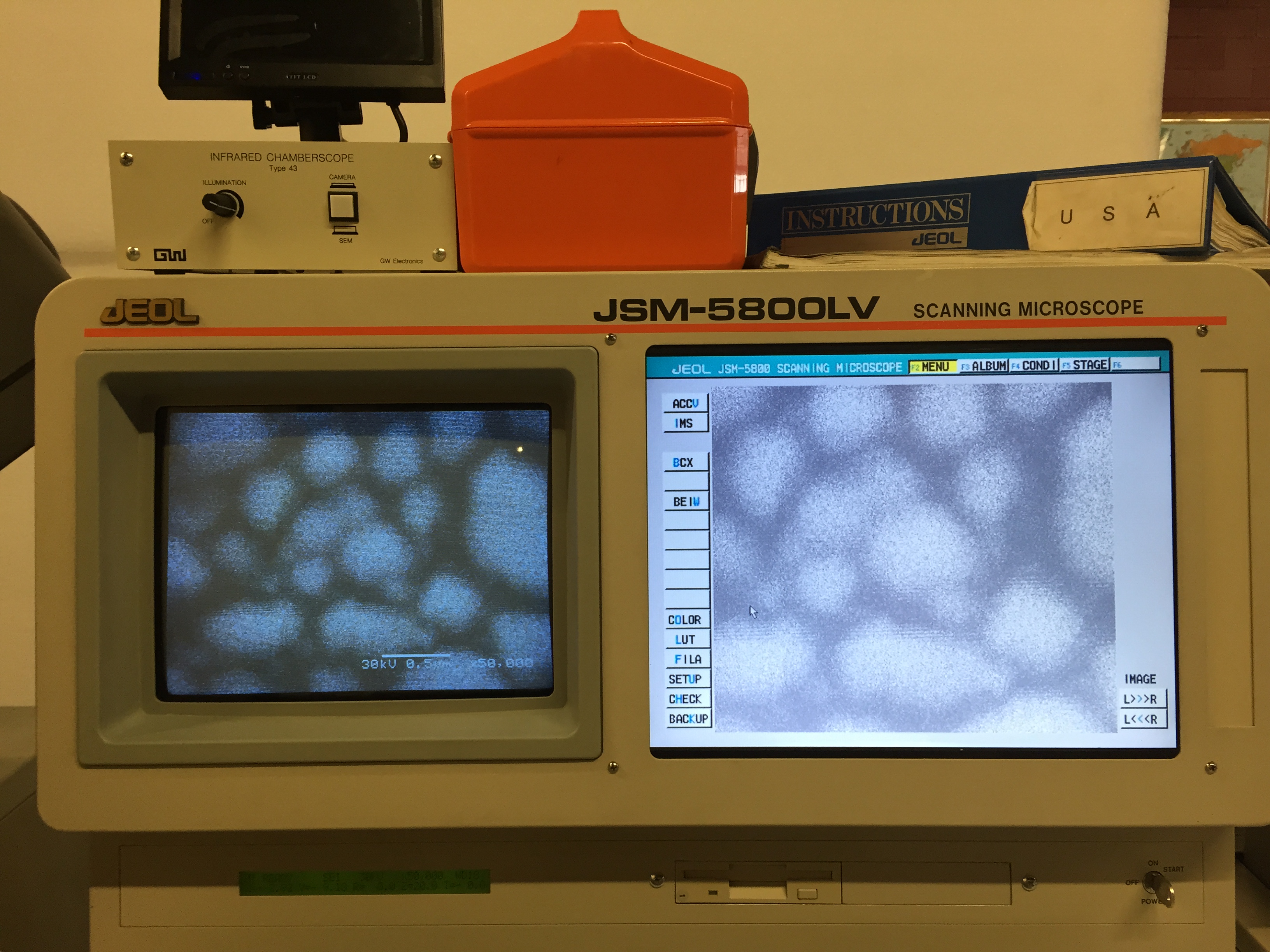
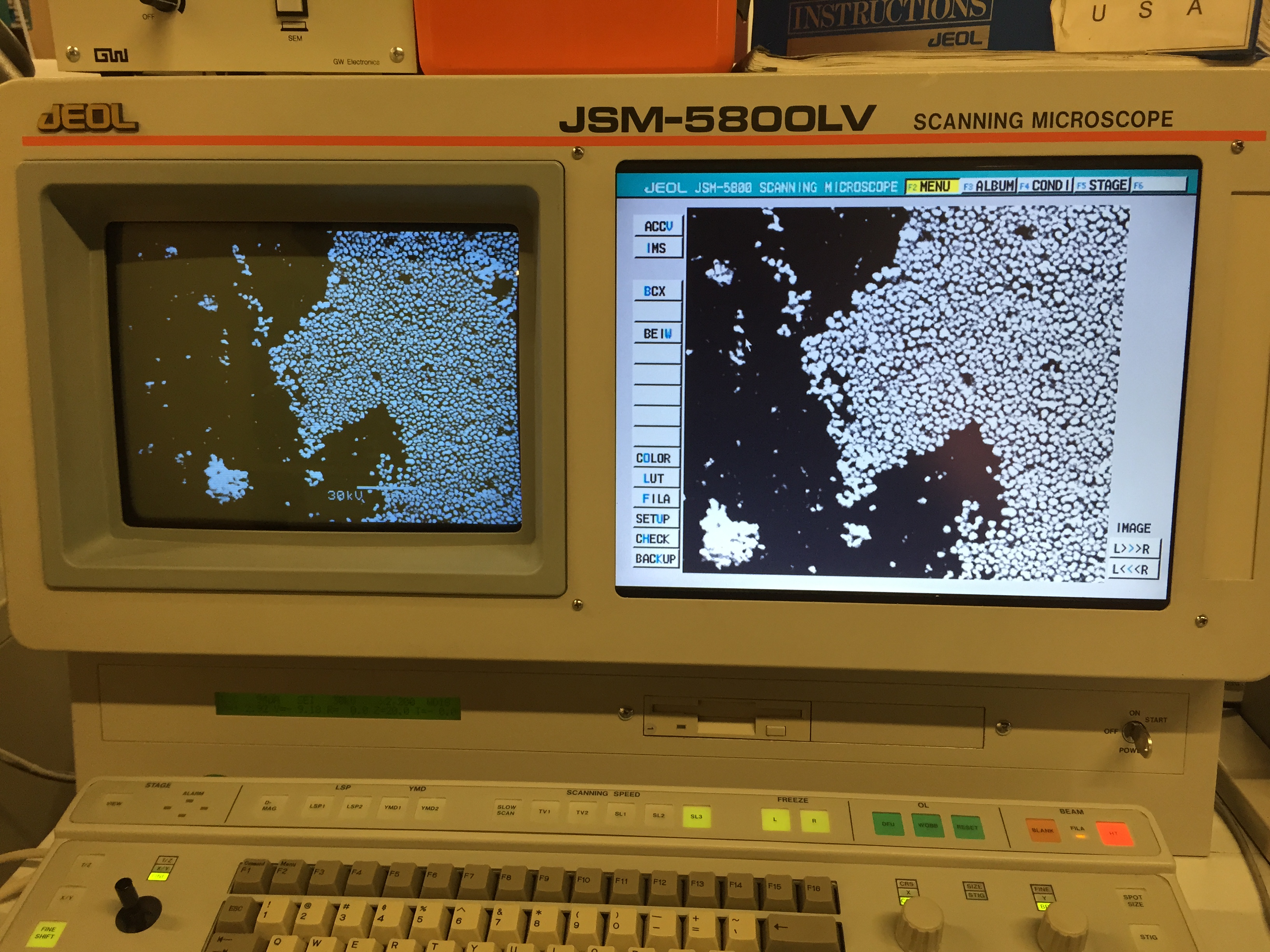
JEOL 5800LV
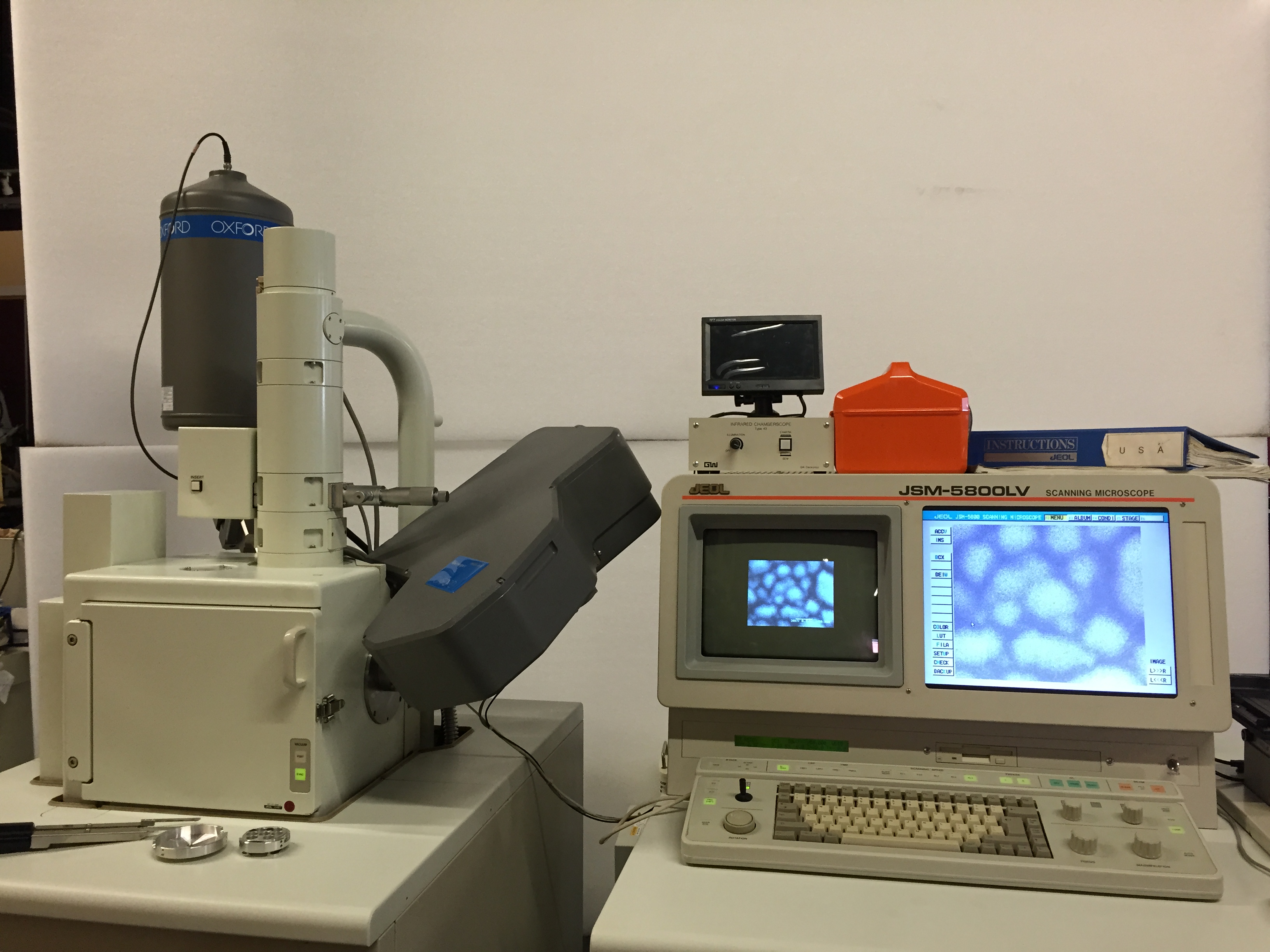
Great entry-level tungsten SEM for the company just getting into running their own samples.
Specifications:
- Fully refurbished, ready for inspection
- 5.5nm resolution in VP mode
- 3.5nm resolution in high vacuum mode
- Up to 300KX magnification
- Solid state backscatter detector
- Tungsten filament
- Digital imaging
- Monitor upgraded to flat-panel
- 100mm x 125mm stage coverage
- Optional EDS and WDS system
Delivery, installation, and training available
Great entry-level tungsten SEM for the company just getting into running their own samples.
Extremely capable for a wide variety of samples, from non-conductive to powders.
Specifications:
- Fully refurbished, ready for inspection
- 5.5nm resolution in VP mode
- 3.5nm resolution in high vacuum mode
- Up to 300KX magnification
- Solid state backscatter detector
- Tungsten filament
- Digital imaging through microscope directly, or through EDS or external capture
- Monitor upgraded to flat-panel
- 100mm x 125mm stage coverage
- Optional EDS and WDS system
Service options available, along with full delivery, installation, and training.